|
KERIC is a core facility offering different imaging analyses in experimental systems in combination with experimental surgery in large animals such as swine as well as in rodents. The unit belongs to the department of comparative medicine (AKM) and is situated at Karolinska University Hospital, Solna. The core facility is financed by Karolinska University Hospital and user fees. We offer a fully equipped facility for experimental surgery with four full size operation units equipped with ventilators and monitors for physiological functions, and two laboratories for small animal surgery. We provide tools for angiography (Philips XD20), MRI (Varian 9.4T), two PET/CT and one PET/MR (Mediso nanoScan), and offer our resources to both academic research, training courses and for external clients to develop and test new equipment.
|
| Hours | Location |
|
Monday - Friday 8:00 - 17:00 |
Karolinska University Hospital, Solna Stockholm, 17176 |
|
Angiography/CT |
iii |
|
|
The unit for angiography provides a monoplane Philips XD20 including 3D angiography and XperCT. It is a joint project with Philips Healthcare offering possibilities for advanced endovascular research, device development and courses in endovascular treatment. The equipment is continuously upgraded and used in activities such as:
|
|
|
|
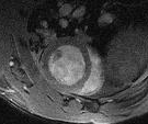
9.4T MRI |
|
|
|
Preclinical MRI equipped with three gradient bore sizes:
There are several coils to cover anatomical imaging of the brain, spine, heart, legs or whole body in various animal models. The system is equipped with four channels and there is also a dedicated Arterial Spin Labelling (ASL) coil for rabbit brain applications. Methods in anatomical imaging, fMRI, pharmacological MRI and cardiac imaging (CINE) are established applications in various small animal models. BIOPAC monitoring system enables following of biological data such as temperature, breathing, heart rate and more. Customized triggering of data scanning to allow images with minimal motion artefacts for breathing or heartbeat. Additional ex-vivo of various sizes with excellent resolution can also be done. |
|
|
|
Experimental Surgery |
|
|
|
Operating rooms for surgical training, courses, and research projects. The four operation units are equipped with:
|
|
|
|
Small animal surgery |
|
|
|
The labs for small animal surgery are equipped with ventilated work areas, isoflurane anaesthesia equipment and a mobile surgical microscope. |
|
|
| Name | Role | Phone | Location |
|---|